Episode 7: Anaphylaxis Part I, Refine your Approach

Episode 7: Anaphylaxis Part I, Refine your Approach

Today, we’ve got not one, but two guest experts to help Dr. Rajiv Thavanathan tackle all things Anaphylaxis! Dr. Graham Wilson is an FRCPC EM resident at The Ottawa Hospital (TOH), who tackled this topic in-detail in his Grand Rounds presentation last year. Joining him is Dr. Derek Lanoue, an incoming McGill Allergy and Immunology Fellow. Let’s dive right in to Part 1 of 2 on this nutty topic!

What is the Diagnostic Criteria of Anaphylaxis?

It’s very difficult to summarize “anaphylaxis” in one definition. To tackle this problem, in 2005, The National Institute of Allergy and Infectious Diseases, and Food Allergy and Anaphylaxis Network came out with the criteria most ED docs probably teach on shift today.

2005 NIAID and FAAN Anaphylaxis Criteria

  • Anaphylaxis is highly likely if:
    • Unknown exposure with acute onset of illness
      • AND Skin OR mucosal changes
      • AND Respiratory compromise OR reduced blood pressure
    • Likely antigen:
      • Two or more organ system involvement: Skin, mucosa, respiratory system, cardiovascular changes, or persistent GI symptoms
    • Known allergen:
      • Reduction in blood pressure

This definition was prospectively validated with a sensitivity 95%, though we aren’t applying it as effectively as it should. A study in the USA by Russell et al. in 2013, and more recently out of the Childrens Hospital of Eastern Ontario in 2020 show that:

  • We are underdiagnosing anaphylaxis upwards of 50% of the time
  • Why? Definitions are too restrictive.
  • “What do persistent GI symptoms even mean?” “Is one episode of emesis enough?”


Things changed for the better in 2020, when the World Allergy Organization tried to simplify things for practitioners. 

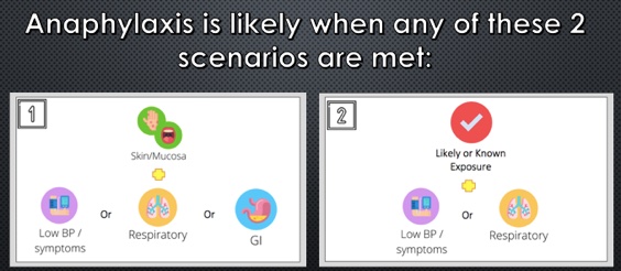
2020 World Allergy Organization Anaphylaxis Criteria

  • Anaphylaxis is highly likely if:
    • Unknown exposure with an acute onset of illness
      • Skin findings, and one other organ system
    • Likely or known antigen
      • Respiratory AND/OR cardiovascular compromise


How can we improve allergy/immunology referrals?

The purpose of our anaphylaxis criteria is to maximize sensitivity to not miss this life-threatening diagnosis, and to administer the antidote, epinephrine, as soon as possible. This poses the question though – what might this approach be sacrificing, from the perspective of the consultant who will see this patient in follow up?

Dr. Lanoue brings up a great point – it’s all in the objective evidence. Did you hear wheezing when you examined them? Do they have hives you can photograph (with consent)? Have they vomited? How many times? Did they have an episode of diarrhea in the ED?

“Okay, but what about tryptase?”

Triptase is an intermediate in anaphylaxis, which causes activation of the compliment system, and mast cell degranulation. This test takes ages to come back, so often it won’t get drawn as part of the work-up for anaphylaxis – some centres won’t have it?

Dr. Lanoue recommends the following from the allergists’ perspective:

  • For the vast majority, get the level.
  • It doesn’t help in the acute setting; it won’t help you decide whether to give epinephrine or not.
  • It can be very useful, however, for the consultant allergist.
  • The level peaks within an hour, and is back to normal within 4-6h. Any patient who presents within this time period from onset may have benefit.

Tryptase levels are useful in the patient where there is an unclear trigger. We know there will be many patients who are tryptase negative, despite being in anaphylaxis. The reverse is also true. In the setting of an unknown trigger, especially when there is diagnostic uncertainty, this may provide another objective piece of evidence for your consultants. Anaphylaxis is not the only diagnosis that is indicated by an elevated tryptase (e.g. mast cell degranulation syndrome).

What is the dosing of epinephrine for anaphylaxis?

  • Epinephrine: 0.01mg/kg, up to a max of 0.5mg.
  • Anyone below 25kg: 0.15mg Epinephrine auto-injector (e.g. Epipen Jr).
  • Anyone above 25kg: 0.3mg Epinephrine auto-injector (e.g. Epipen).
  • We can provide weight-based dosing in the hospital setting, and encourage listeners to do the same to get the most benefit from epinephrine as possible.
  • No established RCT on dosing. Even the 0.01 mg/kg is opinion-based, and based on data on normal adults, not those having anaphylaxis, using measured levels of epinephrine in the blood level.


What is the ideal site of administration for anaphylaxis?

Dr. Estelle Simons is a (Canadian!) prolific researcher in the field of Allergy and anaphylaxis and has served as president of the American Academy of Allergy, Asthma and Immunology (AAAAI). She performed a study measuring peak epinephrine concentrations in healthy, adult volunteers in the deltoid muscle vs the lateral thigh, showing a higher level in the lateral thigh. This is likely due to a higher concentration of vasculature. More of her work showed a difference in the rates of absorption in intramuscular vs subcutaneous administration of epinephrine. Other researchers have demonstrated that the thinner subcutaneous tissue over the lateral thigh allows for more consistent penetration to the muscle.

In summary, use the lateral thigh and use the intramuscular route.

Another huge take away point by both guest speakers is that IV bolus delivery of epinephrine for the treatment of anaphylaxis is associated with increased adverse outcomes compared to both IM and SC.

Use of steroids and antihistamines

These are a very controversial topics, with lots of dogma surrounding the medications we use. In theory, steroids work in two ways. First, it decreases the symptom burden in the acute phase of anaphylaxis. Second, it helps to prevent the concerning, though ever-elusive unicorn that is the biphasic reaction.

In the acute phase, the evidence from steroids came from children with asthma and croup, looking at preventing hospital admissions, and relapse rates. Maximum steroid serum concentrations occur within 1-2 hours, which means that it’s highly unlikely to prevent death from anaphylaxis. We know that death from anaphylaxis occurs within 30 minutes of trigger exposure, which is well outside of the effective window for the steroid.

What we do know is that epinephrine is the first-line intervention for anaphylaxis, with the greatest mortality benefit. Despite, in a cohort study of close to 2000 children diagnosed with anaphylaxis, less than 25% received epinephrine, with almost 50% received steroids.

So what about the biphasic reaction? The biphasic reaction is recurrent anaphylaxis, after complete resolution of symptoms, without re-exposure, within 72 hours of the initial anaphylactic event.

A few facts about the biphasic reaction:

  • Rates range from 4-5% of anaphylaxis cases using the above definition.
  • Most present in a clinically similar manner to the first episode.
  • Only 20% of these patients required additional therapy on top of what they originally received.
  • Steroids in the ED first gained favor in 2007 when a single-centre prospective study showed decreased biphasic reactions with use of steroids.
  • Since that time, several systematic reviews argue against preventative association of biphasic reactions, and one study went so-far as to recommend against the routine use of steroids for anaphylaxis.
  • Further, there have been no reported cases of mortality due to biphasic anaphylaxis as per the 2020 AAAAI Practice Parameters
  • Clinical Features of biphasic anaphylaxis (low-quality evidence):
    • Those who required multiple epinephrine doses
    • Respiratory failure, end-organ damage, cardiovascular shock or arrest.


In summary, don’t extrapolate too much, but the routine use of steroids is probably not ideal. There are, however, select patients that will still benefit:

  • Refractory anaphylaxis
  • Cardiovascular shock
  • Concurrent asthma with anaphylaxis (clear benefit shown with steroids)


“If I’m going to use a steroid, which one should I use?”

If you choose to use a steroid, a careful agent selection that is ideal for your patient can have a much greater benefit than simply throwing IV methylprednisolone at them.

  • Methylprednisolone is associated with the highest rates of steroid-induced anaphylaxis, urticaria, and angioedema of all steroids
  • For this reason, consider using dexamethasone vs hydrocortisone, letting the patients presentation guide our choice
    • When to consider dexamethasone: Dominant upper/lower respiratory involvement are responsive to this medications anti-inflammatory effects, as well as its longer duration of action.
    • When to consider hydrocortisone: If the patient continues to be hypotensive, mineralocorticoid effects from hydrocortisone may play a higher benefit in this shock or shock-like state.

If there’s one take home point from biphasic reactions and steroid use, it’s that the number one medication to prevent relapse and bounce-back from anaphylaxis is epinephrine.

For part 2 episode and show notes, click here !

Episode 7: Anaphylaxis Part I, Refine your Approach

Episode 7: Anaphylaxis Part 2, Ready for Discharge

In part 2 of our 2-part series on Anaphylaxis, our host Dr. Rajiv Thavanathan interviews Dr. Graham Wilson, PGY4 Emergency Medicine at the University of Ottawa and Dr. Derek Lanoue, PGY3 Internal Medicine, University of Ottawa who will soon be an Allergy and Immunology Fellow starting July 1, 2022.

If you haven’t already, you should check-out Part 1 of the series, where the new definition for anaphylaxis and treatment guidelines, including the use of steroids were discussed.

In this part 2, let’s delve deeper into the subject, by discussing monitoring times, discharge instructions and follow-up.

Anaphylaxis Monitoring Times

Although easy to treat with the use of epinephrine, anaphylaxis management is labour intensive in the emergency department with regards to cost effectiveness and human resources, due to the often long “observation” time in the ED. These observation times have a single purpose – monitor for a refractory/biphasic reaction.

Throughout the literature on the topic, there is a wide variation on what is perceived to be an acceptable “mandatory” observation time [range extends between 2-24hrs]. Given the lack of consensus on a set timeframe, some literature has focused on trying to identify this value. A recent study by Ellis has successfully identified that the NPV for the observation of biphasic reaction only increases by 1% for every hour since time of epinephrine injection. This is one of the landmark trials questioning the need for extended observation, primarily for low-risk asymptomatic individuals 1-2hrs post epinephrine injection.

With these promising results, it wouldn’t be inconceivable to think that in the not-so-distant future, risk stratification could be used, and shorter observation periods considered for low-risk individuals. This would certainly help with the human/costly resources needed for extended observation periods.

This conclusion somewhat aligns itself with the 2020 Anaphylaxis guidelines which suggest firmly that high-risk patients require observation for a minimum of 6 hours. These high-risk patients are defined by:

  • Risk Factors: Lack of EMS services, Poor self-management skills, cardiovascular risk factors, and lack of access to epinephrine
  • Requirement for several doses of epinephrine
  • Severe anaphylaxis


Anaphylaxis Mimics: What the ED MD needs to know

Even anaphylaxis has its mimics. These diagnoses are hard to elicit in the moment when a patient presents to the ED with stridor, wheezing and respiratory distress. As such, it’s completely understandable that most ED physicians will provide these patients with at least 1 dose of epinephrine up front. However, if the patient is not responding to epinephrine, there are a few diagnoses that we should perhaps consider.

  • Chronic urticaria [including dermatographism]: This condition, whose pathophysiology is best explained as a hypersensitivity reaction, results in exaggerated weal and flare response to a stimulus – for example physical pressure (being stroked or scrathched) in the case of dermatographism. The condition may respond to the use of antihistamines. It often is an anaphylaxis mimicker when the urticaria is perioral and/or associated with a panic attack, resulting in the physical presentation of tachypnea and dyspnea.
  • Vocal cord dysfunction: This entity is often seen in individuals with allergies to perfumes or other aerosolizing products. In anaphylaxis cross-linking of IgE results in mast cell degranulation. Aromatic rings found in perfumes are incapable of cross-linking IgE or entering the blood stream, and as a result only stimulate a localized hypersensitivity reaction.

In addition to the above mimics, there are several instances when seeing a patient, where the dx of anaphylaxis seems clear from a symptom’s perspective, but the identification of triggers is difficult. It’s important to remember that idiopathic anaphylaxis is a medical entity of its own and is defined by sx of anaphylaxis with no clear trigger.

Furthermore, with the arrival of Lonestar ticks in Canada, Alpha-Gal [sugar molecule found in mammals] allergies from tick bites may gain prevalence. These anaphylactic presentations often are associated with anaphylaxis that wakes a patient up at night, as they encounter a delayed anaphylactic reaction to red meat.

Patient Discharge and Follow-up

Our general threshold for prescribing an epinephrine autoinjector upon discharge should be very low. If you suspect anaphylaxis in any shape or form, it is always safer to simply Rx an autoinjector. Remember that idiopathic anaphylaxis [i.e. anaphylaxis without a clear trigger] is a medical entity, and therefore if you aren’t able to identify a trigger, but their symptoms were consistent with anaphylaxis, you SHOULD prescribe an autoinjector.

Apart from good discharge instructions on when to seek emergency medical attention, how to use their autoinjector safely, a good follow-up is needed for patients presenting with a first-time episode of anaphylaxis. Although human resource constraints for allergist are pronounced in some regions, a referral to a specialist is often beneficial in these individuals. Even if the “trigger” is clear, and the diagnosis of anaphylaxis is clear cut, these patients often have several lingering questions and a need for supports, as this dx will be a “lifelong” struggle. Our allergy specialists can help provide support and reassurance to individuals even if diagnostic clarification is not required.


Take-Home Points 

[TAKE HOME #1] There is ever growing evidence that observation times in the ED can be adjusted based on risk stratification. Although not quite ready for primetime, remember that those with high-risk features [listed below] should be observed for an extended period [i.e. >6hrs].

  • Cardiovascular disease
  • Lack of EMS services
  • Lack of access to epinephrine
  • Poor coping skills

[TAKE HOME #2] Even anaphylaxis has mimics. Consider alternative diagnoses including chronic urticaria and vocal cord dysfunction when epinephrine does not help.

[TAKE HOME #3] Every patient with suspected anaphylaxis should be discharged home with a Rx for an autoinjector, and counselled on it’s use/reasons to return to the ED.

[TAKE HOME #4] Referral to an allergist is not just for dx clarification. It provides an additional line of support for a patient who received an anaphylactic dx that will affect them the rest of their lives.


For more information make sure to check out the EMOttawaBlog post on ANAPHYLAXIS in the ED.


Episode 6: Tick Borne Illness Part 2 – Anaplasmosis

Episode 6: Tick Borne Illness Part 2 – Anaplasmosis

EMOttawa Podcast Episode 6
Tick Borne Illness Part 2 – Anaplasmosis

Click here for Part 1, focusing on Lyme disease.

In part 2 of our 2-part series on tick-borne diseases, Dr. Rajiv Thavanathan interviews Dr. Mikkey McGuinty, an infectious disease clinician-scientist on the topic of anaplasmosis. For a basic review of the disease and pathophysiology, we suggest our previous posts on lyme disease, vector borne illnesses part I and part II. 

Introduction

Anaplasmosis is the broad name for diseases cause by the Gram-negative bacterium Anaplasma phagocytophilumIt is a tick-borne illness, carried by the same Ixodes Scapularis tick that can cause Lyme disease. In humans, it’s called HumanGranulocytic Anaplasmosis (HGA). 

Why are we hearing about Anaplasmosis only recently?

Increased prevalence of ticks with climate change and the evolution of their habitat.

Up until 2020, many physicians had never seen HGA in Ottawa. There were 2 documented cases in Ontario over the last 5-years, both of which were in the Toronto region, which made sense given it’s proximity to the southernmost part of Ontario. Until recently clinicians were not routinely testing for it, and it’s likely that this represents an underestimation of the true prevalence. To date in 2021, there have been 5 documented hospital cases in the Ottawa region 

Epidemiology

  • In Ontario and most of Canada, the same as Lyme disease. That is, tick endemic regions.
    • Ticks in these regions are often found in areas with long grass and low brush. 
    • HGA is more common in Europe. It infects a different tick species (not Ixodes Scapularis), hence the difference in prevalence. 

Classic Presentation

  • Febrile, summer illness.
  • No erythema migrans.
  • Delayed presentation. On-average, illness occurs 1-2 weeks from exposure.
  • Symptoms are general and can include fever, fatigue, myalgias, headache, and less commonly, GI symptoms of nausea, vomiting or diarrhea.

Populations at Risk

  • Elderly (age >60), advanced HIV disease, or impaired T-cell immunity such as organ transplant recipients.
  • Unlike Lyme disease, most people will have a self-limited illness. It doesn’t have as delayed a presentation.
    • However, It can cause severe illness, including syndromes that look like septic shock because of cytokine response to the infection. You can have multi-organ system failure as a result, including brain disease, myocardial involvement, respiratory failure, severe hepatitis, renal failure.
  • 3-5% morbidity, with a case fatality rate of ~1%.

Diagnostic Clues

  • Cytopenia – particularly thrombocytopenia and leukopenia (less commonly anemia)
  • Mild biochemical hepatitis

 Advanced Testing

Bottom line: A challenging diagnosis to make in the ED. Needs to be a clinical diagnosis and confirmatory tests take a while to come back. 

  • Typically, morulae are found in the 1st 3-5 days of the infection, as opposed to later in the course.
  • Serology can be ordered but is only diagnostic when paired. You need an acute sample while sick, and a paired convalescent sample to determine the antigen titre delta.
  • PCR is the gold standard test, and the test of choice in regions with high prevalence.
    • In the ED, this could be helpful for high-suspicion presentations that will be treated empirically or referred to infectious disease.
      • A convincing clinical history, including tick exposure within the last 2-weeks now presenting with fever, myalgias and headache, with thrombocytopenia and biochemical hepatitis.

Prophylaxis & Treatment

  • Does doxycycline prophylaxis change the likelihood of developing HGA?
    • No, at least we don’t have data for it.
    • However, if they have risk factors (RF) for HGA, they have RF for Lyme disease, so if they meet tick criteria for doxycycline prophylaxis this should be given.
  • Treatment is Doxycycline 100mg PO BID x 10-14 days.
    • HGA is incredibly doxycycline sensitive, and the duration could be as short as 7-days, but we’re also concerned about treating Lyme disease.
  • The ID physician’s joke: No one should die without a trial of doxycycline.

Ottawa Specific Testing

At TOH

  • Epic doesn’t have HGA set up as a specific test for blood films
  • If you’re planning to order microscopy and asking to look for morulae, call the hematopathology team to inform them that HGA is on your differential.
    • Simply commenting on this in the EPIC comment section if often missed.

Take-Home Points

  • Anaplasmosis, also known as HGA, is a tick-borne illness carried by the same tick that can cause Lyme disease.
  • The classic presentation is a febrile summer illness, typically 7-days post tick exposure. Symptoms include fever, myalgias, headache, and less commonly, GI symptoms.
  • While typically self-limited, populations at risk of complications are the elderly (age >60), advanced HIV disease, or those with impaired T-cell immunity such as organ transplant recipients.
  • Suggestive lab abnormalities include thrombocytopenia, leukopenia, and mild biochemical hepatitis.
  • Testing in the ED can include ordering microscopy by hematopathology to look for morulae, which are intraneutrophilic bacterial bodies, with peak sensitivity on days 3-5.
  • PCR is the gold-standard test. For patients being treated empirically or referred to infectious disease, this can be considered.
  • Treatment: Doxycycline 100mg PO BID 10-14 days.
  • It’s never the wrong time to offer preventative care for tick-bites.


EMOttawa Podcast Episode 4: Reflections with Ian Stiell

EMOttawa Podcast Episode 4: Reflections with Ian Stiell

Podcast Episode #4 – Dr. Tiffany Lam & Dr. Ian Stiell

(Click here to access Podcast Main menu)

For our fourth episode of the Ottawa EM Podcast, Dr. Tiffany Lam makes her much anticipated Podcast debut! She sits down to talk to Dr. Ian Stiell, a world-renowned clinician and researcher best known for his development of the Ottawa EM decision rules.

How did you get involved in research?

It was ultimately unexpected, as I was not involved in research as a medical student or clinician. It began with leading journal club as a junior staff, learning how studies should be designed, and then going on to pursue a master’s in epidemiology.

Sometimes you need to go with your gut and follow any opportunities that arise, you can’t pre-ordain what’s going to happen.

Who were your mentors growing up?

My father was a forestry researcher, and although not a clinical researcher, I suppose this rubbed off. There was not a lot of emergency medicine research happening early-on in my career, so I relied on a multidisciplinary team of other clinicians early-on.

Advice for students or residents interested in emergency medicine research?

Reach out to known researchers as early as you can. If you’re a medical student, ask for opportunities in your 1st or 2ndyear volunteering or potentially working as a summer student. In Ottawa, there are multiple new EM projects happening each year, including opportunities to get involved helping with the resident research projects.

How does your clinical work influence your research?

After working in the ED for 40-years, it’s really a never-ending opportunity of ideas. Residents are always asking “why do you do it that way?” and you’re expected to know the answer. If you don’t, potentially there’s a new research idea. EM is unique in that we see the early stages of the undifferentiated patient. Whether in identification of disease, risk stratification or prognostication, there are always new opportunities for research in the ED.

What is the most meaningful change you’ve seen regarding the use of evidence in EM?

The shift away from eminence-based medicine, that is, away from old professors telling you what’s right just because it’s always been that way. Now we look at the studies first, and trainees are being taught earlier-on to critically appraise the literature.

How do you maintain a balanced schedule?

Part of it comes with learning how to be more efficient. Working hard, but also being proficient with your time. As an editor-and-chief for CJEM, this includes appraising articles quickly.

How does Ian Stiell prevent burnout?

Right now, it’s through having lots of hobbies outside of medicine and exercising. My hobbies have included skiing, mountain biking, playing golf, tennis and travelling when it’s not a global pandemic. Currently, I’ve been spending a lot of time with my family and grandchildren.

What are some of the greatest life lessons you’ve learned during your career?

You can’t totally preplan your life. In medical school I wanted to be a neurologist, then surgeon, then family doctor, and all of that seemingly went by the wayside as I grew up. You need to be open to new opportunities. Allow yourself to change course if you find something you like better. And don’t be afraid to admit you don’t like the path you’re on and change course.

What’s next for Dr. Ian Stiell?

I’m still actively involved in research, but part of my role has shifted more towards mentoring residents and new researchers. I’m also involved in working as the editor-and-chief of the Canadian Journal of Emergency Medicine (CJEM).

Any final parting words of advice?

While it depends on your personality, emergency medicine is an amazing career to pursue, particularly if you have a short-attention span. It’s exciting, fast paced, a team sport, and there are countless opportunities for clinical research—whether in quality improvement, simulation, or point-of-care ultrasound (POCUS) to name a few.


EMOttawa Podcast Episode 3: Thunderclap Headache

EMOttawa Podcast Episode 3: Thunderclap Headache

On episode three of the Ottawa EM Podcast, Dr. Rajiv Thavanathan (R5) interviews Dr. Michael Hale (R5), on the ED presentation and diagnosis of subarachnoid hemorrhage in the context of the thunderclap headache. (Click here to access Podcast Main menu)

Subarachnoid Hemorrhage (SAH)

Why does the diagnosis matter?

  • 30-day mortality of almost 50%.
  • Of those who survive a SAH diagnosis, 30-50% are left with significant disabilities.
  • While the most common cause of SAH is trauma, this post will specifically focus on non-traumatic SAH, most often due to an aneurysm. 
  • Note while headache account for 2% of all ED visits, SAH accounts for only 1% of this percentile. 
  • Most ER doctors will see less than 50 SAH total during an entire career.

Predictive Signs & Symptoms 

  • Thunderclap headache
  • Worst headache of life
  • Neck stiffness
  • Vomiting
  • Loss of consciousness (LoC)

Note: each of these features alone are more likely to be caused by another diagnosis.

In fact, thunderclap headache, although common in SAH, isn’t overlying predictive of SAH. That is to say, most patients presenting with a describe thunderclap headache will not go on to be diagnosed with a SAH, speaking to the rarity of the diagnosis.

While a combination of the above symptoms increases your pre-test probability, the  combined likelihood still rarely exceeds 25%.

Thunderclap Headache Definitions

  • The inclusion criteria of the Ottawa SAH rule states peaking intensity within 1-hour. This was done to increase the sensitivity of the rule, that is, not to miss any cases.
  • However, the vast majority of SAH peak either instantly, or within a few minutes, very rarely extending beyond minutes.
  • The original textbook definition of thunderclap was “a headache of moderate-to-severe intensity that peaks within 60-seconds”.
  • The story that concerns neurosurgeons “an instantly peaking headache, snap of the fingers fast, comparable to being hit in the head with a baseball bat”.

Non-Contrast CT (NCCT) Head

In 2011, the initial SAH rule-derivation study showed 100% sensitivity to diagnosis SAH if the scan was performed within 6-hours.

However….

In 2020, Dr. Perry’s SAH Validation paper published in Stroke, they found a 95.5% sensitivity. And in-fact the true sensitivity may lie somewhere in-between these numbers, as from the most recent study, of the 5-missed SAH cases of 111 SAH diagnosis:

  • 2 were false positives caused by traumatic lumbar puncture, and the aneurysms identified on CTA were deemed to be incidental by neurosurgery (Note: we know 2-3% of the population will have incidental aneurysms).
  • 1 case missed by radiology.
  • 2 cases were true misses, 1 being a rare cause of SAH (a dural venous fistula), 1 being a patient with sickle-cell anemia and a hemoglobin of only 63 g/L.

*Clinical Pearl: A hemoglobin < 100 g/L makes blood less hyperdense on CT-scan, and therefore much easier to miss. Therefore, clinicians should proceed with caution in applying this rule to the profoundly anemic patient.

For Dr. Michael Hale, if a patient presents to the ED under the 6-hour window, a NCCT will suffice unless the patient is:

  1. Anemic with a hemoglobin of < 100 g/L
  2. Classic story of an instantly peaking headache with neck stiffness, vomiting, where your initial clinical suspicion remains incredibly high.

CT Angiogram (CTA) vs Lumbar Puncture (LP)

  • The literature supports CTA and LP to be effective at ruling out SAH with a similar degree of clinical certainty.
  • For a LP, this does depend on your positive criteria: whether xanthochromia vs. # of RBCs vs. combination of the two.
  • (From Dr. Perry’s study: xanthochromia + 2000 x 106 RBCs as cutoff, combined with a negative NCCT, the miss-rate of SAH is well under 1 in 1000).

Risks:

LP: Pain, bleeding, infection, post-LP headache, time, and can be technically challenging.

CTA: Radiation (double the radiation-dose, albeit still less than that involved a CT Abdo-Pelvis), contrast-allergy, and the risk of detecting an incidental aneurysm (2-3% of the population, the vast majority of which would never cause any problems).

Does time matter for CTA sensitivity as it does for NCCT?

Despite both being CT technology, the two tests are looking for different things. For NCCT, we’re looking for blood in the subarachnoid space, for CTA we’re looking for an aneurysm.

The factors that decrease the sensitivity of each are different:

  • For NCCT, sensitivity is decreased by the amount of blood, the time from onset to time of scan (as blood diffuses away from the area, is broken down and becomes more iso-dense) and anemia.
  • For CTA, sensitivity is decreased by aneurysm location (i.e., near the skull-base, where there is significant artifact), size of the aneurysm, and contrast-timing and technique.
  • Ultimately, what decreases the sensitivity of NCCT notably the timing is very different than what decreases the sensitivity of CTA.

Is there an added diagnostic yield to doing CTA over LP?

  • Yes, when you’re considering alternative diagnosis.
  • i.e., cervical artery dissection, cerebral venous sinus thrombosis (CVST), and reversible cerebral vasoconstriction syndrome (RCVS) can all present with thunderclap headache.

RCVS (Reversible Cerebral Vasoconstrictive Syndrome)

  • First-coined in 2007. A relatively new diagnosis, and more a reflection of recategorization of diagnosis (i.e. the post-coital headache, the exertional thunderclap headache, all likely fall under the category of RCVS).
  • Reversible, segmental vasospasm in the Circle of Willis that’s identified on CTA imaging.
  • These patients often present with recurrent thunderclap headaches i.e. 4 over a 1-month period.
  • This should be on your differential diagnosis in cases of those patients presenting with recurrent thunderclap headache.
  • Although the vast majority of RCVS patients have complete resolution in 1-3 months, a notable percentage of these patients will develop negative sequalae. SAH in 30%, seizures in 15%, and can also lead to stroke and posterior reversible encephalopathy syndrome (PRES).
  • Clinically we can’t predict which RCVS patients will go on to have a benign course, vs. severe complications, therefore neurology often admits these patients for monitoring and initiation or either PO or IV calcium-channel blockers (CCB) to prevent vasospastic complications.

Example

30-year-old patient presenting with thunderclap headache that started 5-hours ago

  • 6-hour NCCT cutoff is sufficient. If normal, stop-there.
  • However, if you’re really considering an alternative diagnosis, i.e. this patient has significant neck pain concerning for cervical artery dissection, or in the immediate post-partum period concerning for CVST, consider proceeding with a CTA even in the below 6-hour group. This isn’t necessarily to rule-out a SAH, but to expand on the differential diagnosis.
  • If the patient with a really concerning story and multiple risk-factors, likely prudent to proceed with LP or CTA even within 6-hours to further lower pre-test probability. *a minority of patients
  • If the hemoglobin is <100g/L, NCCT even within 6-hours is likely non-sufficient.
  • If this patient presented outside the window (i.e., thunderclap headache 8-hours ago), you will want to obtain either a LP or CTA. The decision in choosing between LP and CTA is based both on shared decision-making with the patient on risks vs. benefits, and your clinical suspicion of an alternative thunderclap diagnosis.

Dr. Rajiv Thavanathan sits down with Dr. Jim Yang to talk about some of the best high impact trials in the last year in Emergency Medicine. Which patients with a GI bleed need urgent endoscopy, and which patients ACTUALLY need targeted temperature management (TTM) after cardiac arrest.

Acute Upper GI Bleeds– Does timing matter?

Timing of Endoscopy for Acute Upper Gastrointestinal Bleeding

Design

  • Single centre, RCT performed in Hong Kong
  • 516 patients
  • Patients with overt UGIB, specifically with hematemesis or melena, with a Glasgow Blatchford Score (GBS) of >12

Rapid GBS Review

  • GBS > 1 suggests at risk for needing intervention
  • GBS > 6 traditionally predicted the need for intervention
  • GBS > 12 associated with increased mortality

Intervention

Compared urgent endoscopy (endoscopy performed within 6-hours of GI consult) to early endoscopy (endoscopy performed within 6-24 hours of GI consult). Practically, speaking this means that people in the urgent endoscopy group were scoped overnight if they came in the evening.

All patients received standardized care, consisting of a high-dose PPI, and if concerns for variceal bleeding, vasoactive medications (i.e. octreotide) and antibiotics for spontaneous bacterial peritonitis (SBP) prophylaxis.  

Exclusion Criteria

  • Patients with hypotensive stock who failed to stabilize after initial resuscitation or patients moribund from terminal illness. 

Endoscopic Findings

  • Peptic ulcer disease (PUD) ~60% 
  • Variceal bleeding (7-9% of study)

Note: Epidemiologically, PUD is seen at much higher rates in Asian populations, so this prevalence was expected.  

Outcomes

No difference in all-cause mortality at thirty-days.
No difference in further bleeding, the amount of blood transfusions, the rates of surgery or embolization, or hospital or ICU length of stay. 

Urgent endoscopy is more technically challenging. Waiting gives the medications more times to act, which improves visibility, resulting in less and more-targeted interventions. 

Limitations

Given the study’s low prevalence of variceal bleeds, we can’t apply this to variceal bleeds, a known higher-risk population. This study also excluded patients in shock, so clinical judgement and early involvement of endoscopists is still warranted in these very sick patients. 

Additionally, this only applies to larger centers where you have quick access to endoscopists. Particularly in the community, it’s prudent to still discuss these cases with gastroenterology early, as we know these patients can quickly decompensate.

Case application

60 y/o M, known PUD, CC’ melena & presyncope.
Vitals: HR = 115, BP = 90/60 mmHg
Labs: Hgb = 52 g/L

As per our trial, this is indeed a patient who could be delayed until the next morning for early endoscopy. However, if that patients develops any hemodynamic instability, or signs of ongoing bleeding, Dr. Jim Yang would advocate for an earlier endoscopy.

Therapeutic Hypothermia after ROSC

Is therapeutic hypothermia post ROSC always a good thing?
Or does this depend on the underlying etiology (cardiac vs non-cardiac) and the pre-post ECG rhythm? What temperature should we aim for? 

Pathophysiology Summary:

Anoxic brain injury is associated with increased mortality and morbidity. Fevers are bad and can worsen ischemic brain injury. The theory behind therapeutic hypothermia is that it reduces free radicals, cerebral oxygen consumption, and ultimately reduces ischemic injury.

The initial trials (early 2000s) were small RCTs showing a huge difference in favorable neurologic outcomes for hypothermia post shockable rhythms (post VF and pVT arrest).  

But in 2013 the TTM trial was published: Targeted Temperature Management at 33°C versus 36°C after Cardiac Arrest

The TTM Trial by Nielsen et al. (2013) was a large RCT of 939 patients. It showed that unconscious survivors of out-of-hospital (OOH) cardiac arrest, of presumed cardiac cause (but of all rhythms, shockable and non-shockable) there were no difference in outcomes between a temperature target of 33° C vs. 36° C. 

Flash-forward to the HYPERION Trial this past year: Therapeutic Hypothermia After Cardiac Arrest With Non-Shockable Rhythm

Design

  • Multi-center, RCT performed in 25 ICUs in France
  • 581 adult patients

Included

  • ROSC following in hospital (IH) or out of hospital (OOH) cardiac arrest with non-shockable rhythm due to any cause
  • Patients had to be comatose post (GCS <8)

Excluded

  • Patients with poor prognostic indicators. 
    • More than 10-minutes from collapse to initiation of CPR.
    • More than 60-minutes from initiation of CPR to ROSC
    • Those in refractory shock despite vasopressors

Intervention

  • Randomized to either period of hypothermia (targeted temp 33°) vs normothermia (targeted temp 37°)
  • Hypothermia group was cooled after randomization and maintained at 33° for 24-hours, then slowly rewarmed with fever avoided over the subsequent 24-hours.
  • Normothermia group cooled if need (avoidance of fever) for up to 48-hours

 Outcomes

  • Primary outcome: Survival with favorable neurologic outcome
  • Secondary outcomes: mortality, length of mechanical ventilation, length of ICU stay, adverse events

Results

Hypothermia was associated with improved survival with favourable neurologic outcome. 10% in hypothermia group vs. 6% in normothermia group. However: No difference in overall mortality, no difference in duration of mechanical ventilation, ICU length of stay, or adverse events. 

Note: hypothermia group had TTM for a longer duration than the normothermia group. 

The normothermia had avoidance of fever for 48-hours, whereas the hypothermia group had induction of hypothermia & maintenance for 24 hours, a period of rewarming, and then maintenance of rewarming for an additional 24 hours. In total, between 56-64 hours of TTM. 

Also, a substantial number of patients in the normothermic group were not normothermic! Although target was 37° C, Patients randomized to this group developed fevers > 38° C throughout the TTM period. 

Bottom Line

  1. Fever is bad.
  2. All patients who are comatose following ROSC should be cooled to prevent fever. 
  3. Temperature you end up at isn’t as important as long as you avoid fever. 

For Dr. Jim Yang, the next patient he sees post ROSC, regardless of shockable vs non-shockable rhythm, he’ll be targeting a temperature of 32-36^C, keeping with the American Heart Association and European Resuscitation Counsel.

We should be initiating cooling for all ROSC patients in the ED.

This can be as simply as

  • Inserting a bladder temperature probe
  • Placing ice packs on the patients groin and axilla

 Any regarding rates of adverse events?  

There has never been any document increase in rates of adverse events between hypothermia and normothermia in ANY of published RCT to-date. 

Show notes compiled by Dr. James Gilbertson

Original music by Eusang