Episode 9: Bacterial Peritonitis – Tap that Belly !

Episode 9: Bacterial Peritonitis – Tap that Belly !

In this episode, Dr. Thavanathan interviews Dr. Chirag Bhat, an R5 and chief resident of the Ottawa Emergency Medicine program (at the time of this podcast), on select complications of cirrhosis.

Note that he’s previously written a Grand Rounds Summary of the topic linked here.


For an even deeper dive, we recommend reviewing the 2018 EASL Clinical Practice Guidelines for the management of patients with decompensated cirrhosis.


Patient Case:

Dr. Bhatt highlights a challenging cirrhotic case that identified knowledge gaps. The patient presented with fever, tachycardia, abdominal pain, and had ascites on clinical examination. He suspected spontaneous bacterial peritonitis (SBP) and performed a paracentesis, and the patient met diagnostic criteria for SBP. He prescribed appropriate antibiotics and consulted internal medicine. But while reviewing the chart the following day he learned that despite this, the patient had decompensated. He was later diagnosed with secondary bacterial peritonitis and required emergent surgery.


Biggest Takeaway from the EASL Guidelines:

Cirrhotic patients can have extensive complications from their liver disease. In the emergency department, we often focus on variceal bleeding and less on SBP.  However, SBP has an extremely high mortality and is common. Clinicians should start considering it as the meningitis of the abdomen; in fact, meningitis has a lower mortality rate than SBP. For every four patients that you diagnose with SBP and treat with antibiotics, one will still die in hospital. Of cirrhotic patients, one in four will have at least one episode of SBP during their lifetime. It is both common and deadly.


Diagnosing SBP:

To diagnose SBP, we must do a diagnostic paracentesis and look at the absolute neutrophil count. The paracentesis findings are an ascitic absolute neutrophil count (ANC) of greater than 250 cells/mm.


Is peritonitis needed to diagnose SBP?

No, and in fact this is a common misconception within the name. While we look for fever and abdominal pain, only ½ of patients with SBP present with these symptoms. In fact, fever occurs in only 42% of patients. This is because cirrhotic patients are relatively immunocompromised.


Patients with SBP can present with any of the following:

  • Isolated hepatic encephalopathy
  • Renal failure
  • GI bleeding
  • Any abnormal vital signs
  • Vomiting
  • Metabolic acidosis
  • Leukocytosis


One study, which included paracentesis done in outpatient clinics, even found up to 13% of SBP patients were asymptomatic. The fact that something with a mortality of 25% can be that indolent should terrify you.


If you miss this diagnosis and discharge the patient home, their mortality approaches 90%. That’s why it’s a critical diagnosis. Every hour that you delay a diagnostic paracentesis in a patient with SBP increases their mortality by 3.3%. That’s why this is an ED diagnosis, and why you shouldn’t defer doing the paracentesis to the inpatient team. 


SBP Treatment:

Antibiotics remain a key component of treatment, specifically, a 3rd generation cephalosporin for community acquired SBP in low antibiotic resistance areas. Without antibiotics, SBP has a mortality of 90%. With antibiotics, the mortality is still 25%.


However, there is another extremely important intervention, that can decrease patient mortality from 25% down to 10%. That treatment is albumin.


The EASL guidelines of 2018 recommend that we give albumin at diagnosis at a dose of 1.5 g/kg. This is a grade I recommendation.


It’s based on the theory that albumin would increase intravascular volume but also binds endotoxins in patients with SBP and ultimately prevent hepatorenal syndrome and subsequent death.


This is based on a meta-analysis of four RCT studies done with a total of 288 patients with SBP.

It suggests that albumin has an NNT of 6 to prevent one death, and it has an NNT of 4 to prevent renal failure. So, this is a second critical action you can take on your next shift. Order albumin at 1.5 g/kg within 6 hours of diagnosis for your patients with SBP and help lower the mortality of this dangerous disease.


What is secondary bacterial peritonitis?
About 1 in 20 cases thought to be spontaneous peritonitis are secondary to another source. In contrast to SBP, secondary bacterial peritonitis is when ascites fluid is infected but it’s due to another intraabdominal infection. It’s secondary to another source, like appendicitis, or cholecystitis, or perforation.


When you do the paracentesis, you’ll still have an absolute neutrophil count > 250 cells/mm3, and you will be tempted to call this spontaneous, when in fact, it is secondary from another source. The reason this matters is that secondary peritonitis is a surgical pathology. Without surgery, their mortality is around 100%. Because we wouldn’t be treating the source


How do you differentiate between SBP and Secondary Bacterial Peritonitis?
Unfortunately, this remains a challenge. There’s not much on history that will differentiate between the two. However, on physical exam, secondary bacterial peritonitis patients tend to have more severe and focal abdominal pain.


Is there a clinical tool that can help distinguish between the two?

The Runyon criteria

When you do the paracentesis, you’re looking for 2 of 3 findings in the ascitic fluid:

  1. Protein > 10 g/L
  2. Glucose < 2.8 mmol/L
  3. LDH > ULN of the serum.


These criteria have a specificity of 90%, but a sensitivity of 66% for secondary bacterial peritonitis. So, as emergency physicians, we can’t use Runyon criteria to rule out secondary peritonitis. However, applying the Runyon criteria can push you to do a CT when you weren’t going for other reasons.


Advanced Imaging

Dr. Bhatt highlights a few reasons you should strongly consider performing a CT scan on a patient with cirrhosis and ascites.

  1. Severe abdominal pain
  2. Focal abdominal pain
  3. Positive Runyon criteria
  4. Not responding to typical SBP treatment, including antibiotics and albumin
  5. Also consider a very high ANC as a clue.


Take-home Points:

  • Adopt an extremely low threshold to perform a diagnostic paracentesis in cirrhotic patients. It has an extremely high mortality with a very subtle presentation.

  • The paracentesis findings are an ascitic absolute neutrophil count (ANC) of greater than 250 cells/mm.
  • Only half of SBP patients present with fever and abdominal pain. SBP can present with isolated hepatic encephalopathy, renal failure, GI bleeding, any abnormal vital sign, vomiting, metabolic acidosis or leukocytosis.
  • For SBP treatment, in addition to antibiotics, prescribe albumin at a dose of 1.5 mg/kg as per the latest 2018 EASL guidelines. This is a grade I recommendation.
  • When diagnosing SBP also consider secondary bacterial peritonitis, which occurs in 1 in 20 patients. This is a surgical emergency of the abdomen. Have a low threshold for advanced imaging, particularly in patients with severe or focal abdominal pain, those with meeting Runyon criteria, and patients not responding to treatment as expected.


Episode 8: Patient Complaints

Episode 8: Patient Complaints

On this episode of the EM Ottawa Podcast, Dr. Rajiv Thavanathan talks to  Dr. Nicholas Schouela to discuss the patient complaints process, approaches and mitigation strategies. 


Why do they matter?

They’re common. They’re associated with shame and distress. We’re generally high achieving people who take perceived failures poorly. Importantly, many are based on legitimate patient safety issues. By addressing this, we can work towards improving patient safety.


What has changed recently in the patient safety landscape?

In Canada, between 2016-2020, there was a 20% increase in college complaints across all specialties. The ED sees the 3rd highest rate of college complaints across all specialties; for reference, the highest being obstetrics & gynecology, and the second being general surgery.


How did Covid-19 influence patient complaints?

Despite decreased patient volumes during the initial phases of the pandemic, complaints continued to rise increasing locally in Ottawa by 22%.


What guidelines or legislation exist for hospitals in managing the complaint process?

All provinces have essentially mandated that patient complaints are taken seriously and in a structured way.

Ontario has the Excellent Care for All Act. This is a piece of legislation that was developed to increase the focus and accountability for delivering high-quality care through the initiation of multiple hospital and government level committees.


What is the CMPA, and what is its role in patient complaints?  

The CMPA is a not-for-profit defense association, they provide medical legal advice and assistance, patient compensation, resource for risk management, and they write medical-legal opinions for public policy. For patient complaints, they are there to advise, and if needed provide legal counsel.

What are the steps involved after receiving a patient complaint at the college level?

One of the first steps should involve contacting the CMPA. You should not attempt to access or edit the patient’s chart. Following this, you respond to the complaint with the CMPA’s help. Following your response, the CPSO begins the investigative process; this can take between 3 to 10 months. It then takes 2 to 3 months to formulate a decision; essentially a year-long process.


What are common pitfalls made in responding to patient complaints?

  • Opening the patient’s chart without permission. You need permission from your local hospital to do so as the information does not belong to you.
  • Trying to edit the note or the encounter. If you alter the patient encounter, your credibility is instantly lost.
  • Responding too emotionally or defensively. You want to remain factual and even-toned.


What are the outcomes of complaint decisions?

Split into favorable or unfavorable outcomes. Favorable essentially meaning you were in the right. Unfavorable outcomes include cases that were dismissed with concern, suspensions, restrictions placed on a license, revoking of a license, or voluntary resignation or resignment.


Why do patients complain?

Multiple reasons, although most commonly due to a perceived injustice. A 2018 ED study by the CMPA showed that approximately half of all patient complaints involved scenarios that resulted in patient harm. Of these harm-scenarios, 90% of medical experts were critical of the initial care provided. 

Complaints can be divided into three different categories: Clinical care, situational awareness, or a communication breakdown.


What deficiencies were identified by the CMPA as leading to complaints?

The vast majority pertain to inadequate patient assessments. A good proportion also include failure to refer to appropriate consulting services, and inadequate or incomplete investigations.


What are situational awareness complaints?

On average, 1 out of every 5 complaints are based on situational awareness. These include monitoring issues, such as a patient that should have been on a monitor but was placed in an unmonitored area and harm resulted. This further includes tests that were sent off and not followed up on, patients discharged prior to abnormal lab values resulting, or the physician performing an incomplete review of the medical chart leading to suboptimal care.


What is communication breakdown from the CMPA standpoint?

On average, 1 out of every 4 complaints relate to communication breakdown. These can be divided into four broad categories: physician to patient communication, inter-physician, physician to other allied health providers, and documentation.

Is communication modifiable in reducing patient complaints?

Absolutely. Improving communication can also improve your decision making and situational awareness.


What aspects of handover may reduce patient complaints?

Handover is a high-risk time of physician-to-physician communication breakdown. Poor handover increases the risk of patient harm, time in the department, and investigations.

Dr. Ed Kwok, in a 2020 study identified that ¼ of patients were inadequately handed over, with up to ½ of patients consulted inadequately handed over.

Ultimately, the risks associated with missing key information in handover can be minimized by using a standardized framework, such as ED VITALS.


Beyond a standardized handover framework, how can we further improve communication?

Disclosing non-clinically relevant abnormal results. Particularly if patients have access to reviewing their own labs or electronic charts, as this can be distressing to patients. Addressing all incidental findings on imaging, even if it simply means following up with their primary care provider in 6-months to repeat the test.


Tips for disclosing errors

Firstly, certain situations mandate the disclosure of error. This includes any incident that leads directly to patient-harm, or that reached the patient and had the potential for harm, even if it didn’t cause any harm. Generally, it’s a good idea to disclose any incident that reaches the patient even if it didn’t have the potential for harm.


Any pearls or pitfalls for apologizing to patients?

An apology in the form of a statement of regret is both a good idea and can help with emotional healing. This is protected across Canada in different provincial acts as not being considered an admission of fault. For reference, in Ontario, this act is called the Apology Act. 


What are the critical elements of discharge instructions that can minimize patient complaints?

This is the final impression you give to a patient and relays critical information. You should include advise specific to their condition, a follow-up plan, the signs and symptoms in which you would want them to seek care again and make it clear that they are always welcome to return. This should be done verbally at a minimum; however, written handouts can be a nice compliment to this.


Where do we go wrong with obtaining and documenting consent in the ED?

Two different categories. The first is forgetting to obtain consent entirely, which can lead to allegations of battery. The second is a consent process that fails to adequately inform them. Recall that consent includes multiple components: it needs to be voluntary, the patient needs to have capacity, they need to be properly informed about the risks, benefits, alternatives, and consequences of declining the procedure.


In the age of electronic charting, what are the medicolegal risks of using pre-filled templates or dot-phrases?

While largely beneficial, they can lead you to documenting things you didn’t do, which can of course harm you medicolegally. There hasn’t been a specific CMPA judgement on this that the authors are aware of.


Any advice to be more empathetic or caring for patients in the emergency department?

Remember that patients present to the ED because they have a worry or concern. Even if they don’t have underlying emergent pathology, they don’t feel well. Using empathy to address these concerns remains paramount. Additionally, use a patient-centered approach with culturally appropriate care. 


What practical advice do you have for physicians dealing with patient complaints?

Recognize that these aren’t easy. We will all receive at least one during our career no matter how careful we are. We know that complaints can cause significant emotional distress, anxiety, depression, substance abuse, social withdrawal, work disruptions, insomnia, loss of trust in patients, and feelings of helplessness. Self-care is important: exercise, mindfulness, and seeking out supportive people in your network or professional support. There are provincial medical associations physician help programs. If you work out of a university, there are also university affiliated faculty wellness programs. The CMPA itself also has several resources and guides.


Any practical strategies that can be exercised independently?

Cognitive strategies, such as the best-friend strategy, which involves treating yourself how you would treat your best friend if they came to you in the same scenario. Reflect on maintaining a growth mindsight and consider reviewing your knowledge base if there were any identified areas of deficit. Most importantly, practicing self-care as emphasized above.


Episode 7: Anaphylaxis Part I, Refine your Approach

Episode 7: Anaphylaxis Part I, Refine your Approach

Today, we’ve got not one, but two guest experts to help Dr. Rajiv Thavanathan tackle all things Anaphylaxis! Dr. Graham Wilson is an FRCPC EM resident at The Ottawa Hospital (TOH), who tackled this topic in-detail in his Grand Rounds presentation last year. Joining him is Dr. Derek Lanoue, an incoming McGill Allergy and Immunology Fellow. Let’s dive right in to Part 1 of 2 on this nutty topic!

What is the Diagnostic Criteria of Anaphylaxis?

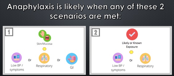
It’s very difficult to summarize “anaphylaxis” in one definition. To tackle this problem, in 2005, The National Institute of Allergy and Infectious Diseases, and Food Allergy and Anaphylaxis Network came out with the criteria most ED docs probably teach on shift today.

2005 NIAID and FAAN Anaphylaxis Criteria

  • Anaphylaxis is highly likely if:
    • Unknown exposure with acute onset of illness
      • AND Skin OR mucosal changes
      • AND Respiratory compromise OR reduced blood pressure
    • Likely antigen:
      • Two or more organ system involvement: Skin, mucosa, respiratory system, cardiovascular changes, or persistent GI symptoms
    • Known allergen:
      • Reduction in blood pressure

This definition was prospectively validated with a sensitivity 95%, though we aren’t applying it as effectively as it should. A study in the USA by Russell et al. in 2013, and more recently out of the Childrens Hospital of Eastern Ontario in 2020 show that:

  • We are underdiagnosing anaphylaxis upwards of 50% of the time
  • Why? Definitions are too restrictive.
  • “What do persistent GI symptoms even mean?” “Is one episode of emesis enough?”


Things changed for the better in 2020, when the World Allergy Organization tried to simplify things for practitioners. 

2020 World Allergy Organization Anaphylaxis Criteria

  • Anaphylaxis is highly likely if:
    • Unknown exposure with an acute onset of illness
      • Skin findings, and one other organ system
    • Likely or known antigen
      • Respiratory AND/OR cardiovascular compromise


How can we improve allergy/immunology referrals?

The purpose of our anaphylaxis criteria is to maximize sensitivity to not miss this life-threatening diagnosis, and to administer the antidote, epinephrine, as soon as possible. This poses the question though – what might this approach be sacrificing, from the perspective of the consultant who will see this patient in follow up?

Dr. Lanoue brings up a great point – it’s all in the objective evidence. Did you hear wheezing when you examined them? Do they have hives you can photograph (with consent)? Have they vomited? How many times? Did they have an episode of diarrhea in the ED?

“Okay, but what about tryptase?”

Triptase is an intermediate in anaphylaxis, which causes activation of the compliment system, and mast cell degranulation. This test takes ages to come back, so often it won’t get drawn as part of the work-up for anaphylaxis – some centres won’t have it?

Dr. Lanoue recommends the following from the allergists’ perspective:

  • For the vast majority, get the level.
  • It doesn’t help in the acute setting; it won’t help you decide whether to give epinephrine or not.
  • It can be very useful, however, for the consultant allergist.
  • The level peaks within an hour, and is back to normal within 4-6h. Any patient who presents within this time period from onset may have benefit.

Tryptase levels are useful in the patient where there is an unclear trigger. We know there will be many patients who are tryptase negative, despite being in anaphylaxis. The reverse is also true. In the setting of an unknown trigger, especially when there is diagnostic uncertainty, this may provide another objective piece of evidence for your consultants. Anaphylaxis is not the only diagnosis that is indicated by an elevated tryptase (e.g. mast cell degranulation syndrome).

What is the dosing of epinephrine for anaphylaxis?

  • Epinephrine: 0.01mg/kg, up to a max of 0.5mg.
  • Anyone below 25kg: 0.15mg Epinephrine auto-injector (e.g. Epipen Jr).
  • Anyone above 25kg: 0.3mg Epinephrine auto-injector (e.g. Epipen).
  • We can provide weight-based dosing in the hospital setting, and encourage listeners to do the same to get the most benefit from epinephrine as possible.
  • No established RCT on dosing. Even the 0.01 mg/kg is opinion-based, and based on data on normal adults, not those having anaphylaxis, using measured levels of epinephrine in the blood level.


What is the ideal site of administration for anaphylaxis?

Dr. Estelle Simons is a (Canadian!) prolific researcher in the field of Allergy and anaphylaxis and has served as president of the American Academy of Allergy, Asthma and Immunology (AAAAI). She performed a study measuring peak epinephrine concentrations in healthy, adult volunteers in the deltoid muscle vs the lateral thigh, showing a higher level in the lateral thigh. This is likely due to a higher concentration of vasculature. More of her work showed a difference in the rates of absorption in intramuscular vs subcutaneous administration of epinephrine. Other researchers have demonstrated that the thinner subcutaneous tissue over the lateral thigh allows for more consistent penetration to the muscle.

In summary, use the lateral thigh and use the intramuscular route.

Another huge take away point by both guest speakers is that IV bolus delivery of epinephrine for the treatment of anaphylaxis is associated with increased adverse outcomes compared to both IM and SC.

Use of steroids and antihistamines

These are a very controversial topics, with lots of dogma surrounding the medications we use. In theory, steroids work in two ways. First, it decreases the symptom burden in the acute phase of anaphylaxis. Second, it helps to prevent the concerning, though ever-elusive unicorn that is the biphasic reaction.

In the acute phase, the evidence from steroids came from children with asthma and croup, looking at preventing hospital admissions, and relapse rates. Maximum steroid serum concentrations occur within 1-2 hours, which means that it’s highly unlikely to prevent death from anaphylaxis. We know that death from anaphylaxis occurs within 30 minutes of trigger exposure, which is well outside of the effective window for the steroid.

What we do know is that epinephrine is the first-line intervention for anaphylaxis, with the greatest mortality benefit. Despite, in a cohort study of close to 2000 children diagnosed with anaphylaxis, less than 25% received epinephrine, with almost 50% received steroids.

So what about the biphasic reaction? The biphasic reaction is recurrent anaphylaxis, after complete resolution of symptoms, without re-exposure, within 72 hours of the initial anaphylactic event.

A few facts about the biphasic reaction:

  • Rates range from 4-5% of anaphylaxis cases using the above definition.
  • Most present in a clinically similar manner to the first episode.
  • Only 20% of these patients required additional therapy on top of what they originally received.
  • Steroids in the ED first gained favor in 2007 when a single-centre prospective study showed decreased biphasic reactions with use of steroids.
  • Since that time, several systematic reviews argue against preventative association of biphasic reactions, and one study went so-far as to recommend against the routine use of steroids for anaphylaxis.
  • Further, there have been no reported cases of mortality due to biphasic anaphylaxis as per the 2020 AAAAI Practice Parameters
  • Clinical Features of biphasic anaphylaxis (low-quality evidence):
    • Those who required multiple epinephrine doses
    • Respiratory failure, end-organ damage, cardiovascular shock or arrest.


In summary, don’t extrapolate too much, but the routine use of steroids is probably not ideal. There are, however, select patients that will still benefit:

  • Refractory anaphylaxis
  • Cardiovascular shock
  • Concurrent asthma with anaphylaxis (clear benefit shown with steroids)


“If I’m going to use a steroid, which one should I use?”

If you choose to use a steroid, a careful agent selection that is ideal for your patient can have a much greater benefit than simply throwing IV methylprednisolone at them.

  • Methylprednisolone is associated with the highest rates of steroid-induced anaphylaxis, urticaria, and angioedema of all steroids
  • For this reason, consider using dexamethasone vs hydrocortisone, letting the patients presentation guide our choice
    • When to consider dexamethasone: Dominant upper/lower respiratory involvement are responsive to this medications anti-inflammatory effects, as well as its longer duration of action.
    • When to consider hydrocortisone: If the patient continues to be hypotensive, mineralocorticoid effects from hydrocortisone may play a higher benefit in this shock or shock-like state.

If there’s one take home point from biphasic reactions and steroid use, it’s that the number one medication to prevent relapse and bounce-back from anaphylaxis is epinephrine.

For part 2 episode and show notes, click here !

Episode 7: Anaphylaxis Part I, Refine your Approach

Episode 7: Anaphylaxis Part 2, Ready for Discharge

In part 2 of our 2-part series on Anaphylaxis, our host Dr. Rajiv Thavanathan interviews Dr. Graham Wilson, PGY4 Emergency Medicine at the University of Ottawa and Dr. Derek Lanoue, PGY3 Internal Medicine, University of Ottawa who will soon be an Allergy and Immunology Fellow starting July 1, 2022.

If you haven’t already, you should check-out Part 1 of the series, where the new definition for anaphylaxis and treatment guidelines, including the use of steroids were discussed.

In this part 2, let’s delve deeper into the subject, by discussing monitoring times, discharge instructions and follow-up.

Anaphylaxis Monitoring Times

Although easy to treat with the use of epinephrine, anaphylaxis management is labour intensive in the emergency department with regards to cost effectiveness and human resources, due to the often long “observation” time in the ED. These observation times have a single purpose – monitor for a refractory/biphasic reaction.

Throughout the literature on the topic, there is a wide variation on what is perceived to be an acceptable “mandatory” observation time [range extends between 2-24hrs]. Given the lack of consensus on a set timeframe, some literature has focused on trying to identify this value. A recent study by Ellis has successfully identified that the NPV for the observation of biphasic reaction only increases by 1% for every hour since time of epinephrine injection. This is one of the landmark trials questioning the need for extended observation, primarily for low-risk asymptomatic individuals 1-2hrs post epinephrine injection.

With these promising results, it wouldn’t be inconceivable to think that in the not-so-distant future, risk stratification could be used, and shorter observation periods considered for low-risk individuals. This would certainly help with the human/costly resources needed for extended observation periods.

This conclusion somewhat aligns itself with the 2020 Anaphylaxis guidelines which suggest firmly that high-risk patients require observation for a minimum of 6 hours. These high-risk patients are defined by:

  • Risk Factors: Lack of EMS services, Poor self-management skills, cardiovascular risk factors, and lack of access to epinephrine
  • Requirement for several doses of epinephrine
  • Severe anaphylaxis


Anaphylaxis Mimics: What the ED MD needs to know

Even anaphylaxis has its mimics. These diagnoses are hard to elicit in the moment when a patient presents to the ED with stridor, wheezing and respiratory distress. As such, it’s completely understandable that most ED physicians will provide these patients with at least 1 dose of epinephrine up front. However, if the patient is not responding to epinephrine, there are a few diagnoses that we should perhaps consider.

  • Chronic urticaria [including dermatographism]: This condition, whose pathophysiology is best explained as a hypersensitivity reaction, results in exaggerated weal and flare response to a stimulus – for example physical pressure (being stroked or scrathched) in the case of dermatographism. The condition may respond to the use of antihistamines. It often is an anaphylaxis mimicker when the urticaria is perioral and/or associated with a panic attack, resulting in the physical presentation of tachypnea and dyspnea.
  • Vocal cord dysfunction: This entity is often seen in individuals with allergies to perfumes or other aerosolizing products. In anaphylaxis cross-linking of IgE results in mast cell degranulation. Aromatic rings found in perfumes are incapable of cross-linking IgE or entering the blood stream, and as a result only stimulate a localized hypersensitivity reaction.

In addition to the above mimics, there are several instances when seeing a patient, where the dx of anaphylaxis seems clear from a symptom’s perspective, but the identification of triggers is difficult. It’s important to remember that idiopathic anaphylaxis is a medical entity of its own and is defined by sx of anaphylaxis with no clear trigger.

Furthermore, with the arrival of Lonestar ticks in Canada, Alpha-Gal [sugar molecule found in mammals] allergies from tick bites may gain prevalence. These anaphylactic presentations often are associated with anaphylaxis that wakes a patient up at night, as they encounter a delayed anaphylactic reaction to red meat.

Patient Discharge and Follow-up

Our general threshold for prescribing an epinephrine autoinjector upon discharge should be very low. If you suspect anaphylaxis in any shape or form, it is always safer to simply Rx an autoinjector. Remember that idiopathic anaphylaxis [i.e. anaphylaxis without a clear trigger] is a medical entity, and therefore if you aren’t able to identify a trigger, but their symptoms were consistent with anaphylaxis, you SHOULD prescribe an autoinjector.

Apart from good discharge instructions on when to seek emergency medical attention, how to use their autoinjector safely, a good follow-up is needed for patients presenting with a first-time episode of anaphylaxis. Although human resource constraints for allergist are pronounced in some regions, a referral to a specialist is often beneficial in these individuals. Even if the “trigger” is clear, and the diagnosis of anaphylaxis is clear cut, these patients often have several lingering questions and a need for supports, as this dx will be a “lifelong” struggle. Our allergy specialists can help provide support and reassurance to individuals even if diagnostic clarification is not required.


Take-Home Points 

[TAKE HOME #1] There is ever growing evidence that observation times in the ED can be adjusted based on risk stratification. Although not quite ready for primetime, remember that those with high-risk features [listed below] should be observed for an extended period [i.e. >6hrs].

  • Cardiovascular disease
  • Lack of EMS services
  • Lack of access to epinephrine
  • Poor coping skills

[TAKE HOME #2] Even anaphylaxis has mimics. Consider alternative diagnoses including chronic urticaria and vocal cord dysfunction when epinephrine does not help.

[TAKE HOME #3] Every patient with suspected anaphylaxis should be discharged home with a Rx for an autoinjector, and counselled on it’s use/reasons to return to the ED.

[TAKE HOME #4] Referral to an allergist is not just for dx clarification. It provides an additional line of support for a patient who received an anaphylactic dx that will affect them the rest of their lives.


For more information make sure to check out the EMOttawaBlog post on ANAPHYLAXIS in the ED.


Episode 6: Tick Borne Illness Part 2 – Anaplasmosis

Episode 6: Tick Borne Illness Part 2 – Anaplasmosis

EMOttawa Podcast Episode 6
Tick Borne Illness Part 2 – Anaplasmosis

Click here for Part 1, focusing on Lyme disease.

In part 2 of our 2-part series on tick-borne diseases, Dr. Rajiv Thavanathan interviews Dr. Mikkey McGuinty, an infectious disease clinician-scientist on the topic of anaplasmosis. For a basic review of the disease and pathophysiology, we suggest our previous posts on lyme disease, vector borne illnesses part I and part II. 

Introduction

Anaplasmosis is the broad name for diseases cause by the Gram-negative bacterium Anaplasma phagocytophilumIt is a tick-borne illness, carried by the same Ixodes Scapularis tick that can cause Lyme disease. In humans, it’s called HumanGranulocytic Anaplasmosis (HGA). 

Why are we hearing about Anaplasmosis only recently?

Increased prevalence of ticks with climate change and the evolution of their habitat.

Up until 2020, many physicians had never seen HGA in Ottawa. There were 2 documented cases in Ontario over the last 5-years, both of which were in the Toronto region, which made sense given it’s proximity to the southernmost part of Ontario. Until recently clinicians were not routinely testing for it, and it’s likely that this represents an underestimation of the true prevalence. To date in 2021, there have been 5 documented hospital cases in the Ottawa region 

Epidemiology

  • In Ontario and most of Canada, the same as Lyme disease. That is, tick endemic regions.
    • Ticks in these regions are often found in areas with long grass and low brush. 
    • HGA is more common in Europe. It infects a different tick species (not Ixodes Scapularis), hence the difference in prevalence. 

Classic Presentation

  • Febrile, summer illness.
  • No erythema migrans.
  • Delayed presentation. On-average, illness occurs 1-2 weeks from exposure.
  • Symptoms are general and can include fever, fatigue, myalgias, headache, and less commonly, GI symptoms of nausea, vomiting or diarrhea.

Populations at Risk

  • Elderly (age >60), advanced HIV disease, or impaired T-cell immunity such as organ transplant recipients.
  • Unlike Lyme disease, most people will have a self-limited illness. It doesn’t have as delayed a presentation.
    • However, It can cause severe illness, including syndromes that look like septic shock because of cytokine response to the infection. You can have multi-organ system failure as a result, including brain disease, myocardial involvement, respiratory failure, severe hepatitis, renal failure.
  • 3-5% morbidity, with a case fatality rate of ~1%.

Diagnostic Clues

  • Cytopenia – particularly thrombocytopenia and leukopenia (less commonly anemia)
  • Mild biochemical hepatitis

 Advanced Testing

Bottom line: A challenging diagnosis to make in the ED. Needs to be a clinical diagnosis and confirmatory tests take a while to come back. 

  • Typically, morulae are found in the 1st 3-5 days of the infection, as opposed to later in the course.
  • Serology can be ordered but is only diagnostic when paired. You need an acute sample while sick, and a paired convalescent sample to determine the antigen titre delta.
  • PCR is the gold standard test, and the test of choice in regions with high prevalence.
    • In the ED, this could be helpful for high-suspicion presentations that will be treated empirically or referred to infectious disease.
      • A convincing clinical history, including tick exposure within the last 2-weeks now presenting with fever, myalgias and headache, with thrombocytopenia and biochemical hepatitis.

Prophylaxis & Treatment

  • Does doxycycline prophylaxis change the likelihood of developing HGA?
    • No, at least we don’t have data for it.
    • However, if they have risk factors (RF) for HGA, they have RF for Lyme disease, so if they meet tick criteria for doxycycline prophylaxis this should be given.
  • Treatment is Doxycycline 100mg PO BID x 10-14 days.
    • HGA is incredibly doxycycline sensitive, and the duration could be as short as 7-days, but we’re also concerned about treating Lyme disease.
  • The ID physician’s joke: No one should die without a trial of doxycycline.

Ottawa Specific Testing

At TOH

  • Epic doesn’t have HGA set up as a specific test for blood films
  • If you’re planning to order microscopy and asking to look for morulae, call the hematopathology team to inform them that HGA is on your differential.
    • Simply commenting on this in the EPIC comment section if often missed.

Take-Home Points

  • Anaplasmosis, also known as HGA, is a tick-borne illness carried by the same tick that can cause Lyme disease.
  • The classic presentation is a febrile summer illness, typically 7-days post tick exposure. Symptoms include fever, myalgias, headache, and less commonly, GI symptoms.
  • While typically self-limited, populations at risk of complications are the elderly (age >60), advanced HIV disease, or those with impaired T-cell immunity such as organ transplant recipients.
  • Suggestive lab abnormalities include thrombocytopenia, leukopenia, and mild biochemical hepatitis.
  • Testing in the ED can include ordering microscopy by hematopathology to look for morulae, which are intraneutrophilic bacterial bodies, with peak sensitivity on days 3-5.
  • PCR is the gold-standard test. For patients being treated empirically or referred to infectious disease, this can be considered.
  • Treatment: Doxycycline 100mg PO BID 10-14 days.
  • It’s never the wrong time to offer preventative care for tick-bites.